More uses have been found for tagging cells with nano-particles, says S.Ananthanarayanan.
Just a few months ago, the journal, Nature Nano-technology carried a report of getting nano-particles which are embedded in living cells to glow strongly so that single cells could be made out. Some members of the same group of scientists, with others, now report an improvement, to get different nano-particles to flash at different times after excitation, so that they could be told apart. This is a feature that can be used in many fields - to mark and monitor different kinds of individual cells, in creating fine grained data storage or even multiplying the unique features of documents like currency notes or credit cards, to discourage counterfeits.
Yiqing Lu, Jiangbo Zhao, Run Zhang, Yujia Liu, Deming Liu, Ewa M. Goldys, Xusan Yang, Peng Xi, Anwar Sunna, Jie Lu, Yu Shi, Robert C. Leif, Yujing Huo, Jian Shen, James A. Piper, J. Paul Robinson and Dayong Jin, in Autralia, Beijing, Shanghai, California and Indiana, report in the journal, Nature Photonics that they have been able to tune the time it takes for nano-particles to decay, that is, to emit light after excitation, to vary from 25 microseconds to 662 micrseconds. Differently tuned nano-particles would then glow after their individual decay times and allow observers to tell them apart.
The technique is one way of using the same medium to carry more than one message. The idea has been refined in the field of telephone communication, where a single copper wire carries currents alternating at different frequencies. The way speech, or even data, is transmitted is by loading the signal on to a high frequency carrier wave, either over a copper wire or even a radio wave. Different streams of voice or data are loaded on different frequencies and sent out together. Receivers can then separate the different carriers, using frequency filters, and detect the different messages. The technique is called multiplexing and is routine in telecommunication, with huge numbers of frequencies loaded on a single optical fibre cable, for instance. And there are different kinds of multiplexing.
In the life sciences, the idea of multiplexing is used for detecting and counting members of different species at the same time. To work out the genetic features of a person’s DNA, to create a tailor-made therapy for that person, for instance, each genetic feature would need to be identified in a series of scans of the DNA. But if the different features of interest could be differently labeled, the whole task could be done in a single scan. In data storage, the limitation is the number of storage elements in a given space. Now, if each element were capable of carrying more than one kind of mark, then that many different sets of data could be stored using the same number of elements. In security printing of documents, a device is to add features over and above the simple printed matter – some mark that glows in ultra violet light, for example. A genuine document can then be identified by viewing in UV light – and the more such features there are, the greater the security.
“Multiplexing typically requires a matrix of optical codes, ideally carried by nano or micro-sized objects, each of which should be accurately identifiable at high speed and at low cost,” say the authors in the paper. A useful way of multiplexing is fluorescenc, a phenomenon where atoms in materials are excited by absorption of light of a particular colour and then emit light, usually at a lower frequency. The best known example is the fluorescent lamp, or the tube light, where the coating on the lamp absorbs ultra violet light and gives off nearly white light. Using different fluorescent markers has been a popular way of multiplexing in detecting cells in biology, since some decades. But the method has the limitation, the authors note, of not more than about 20 colours being clearly usable, and the method calls for three to five lasers, a large number of filters and as many detectors as colours. There is hence the need for other means of coding biological tissue and different kinds of spectral analyses and even time delays in fluorescence are being made use of.
A problem in these methods, of getting target objects to give off different unique signals, is that the glare of the original, incident radiation, obscures the faint light that has to be detected. One way out has been through the phenomenon of upconversion, which is where nano-particles absorb more than one photon of the incident light and then emit at a higher, as opposed to a lower frequency. In this way, the original light can be in the infra-red but the emitted light in the visible region, which would eliminate visible glare. The work that the multinational group had last reported was to engineer the nano-particles to give off a strong enough unconverted signal to be useful.
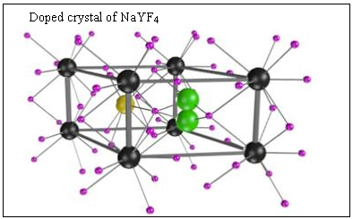
In the current paper, the group reports a way that upconverted light can provide a further dimension of multiplexing. The manner of using upconversion nano-particles has been with crystals of sodium-yttrium flouride (NaYF4) which have been doped with atoms of ytterbium and thulium. Ytterbium is the sensitizer, which absorbs a photon of light and transfers energy, without radiation, to thulium atom emitters. While the earlier work was to find optimum concentration of sensitizer and emitter, with strong excitation radiation, for the strongest emitted signal, the current work has been to modify the sensitiemitter concentrations to vary the distance between the two kinds of atoms in structure of the NaYF4 crystal, and hence to vary the time between excitation and emission.
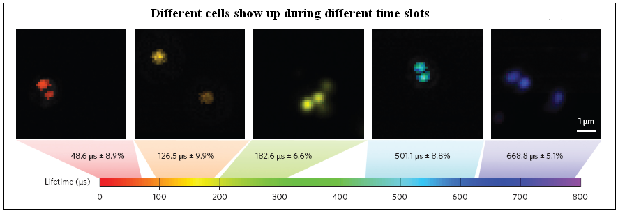
The group reports nano-particles that emit blue light with delays of a wide range, from 48 microseconds to 668 microseconds. Different kinds of cells can then be embedded with nano-particles that have different delays, and then detected, not by the different colours emitted, but by the emissions at different times after the excitation. The detection during separate time slots also serves to eliminate background light and the method has proved to be sensitive and capable of single nano-particle detection.
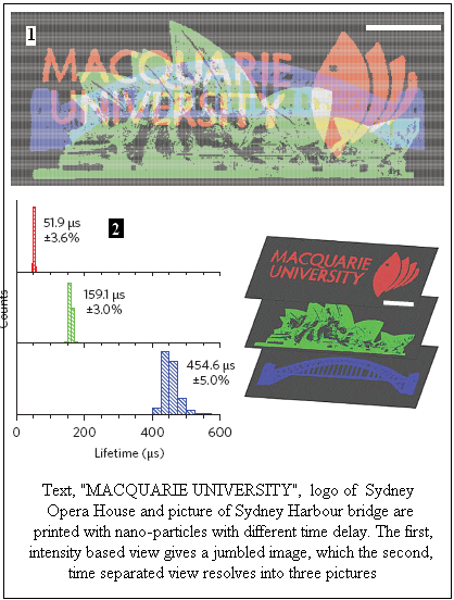
The first picture shows samples of different kinds of cells detected with the help of nano-particles that emit with different delay. The second picture is of three images embedded in the same document, and each image becoming visible according to the time window in which the document is viewed.
------------------------------------------------------------------------------------------ Do respond to : response@simplescience.in------------------------------------------- 